
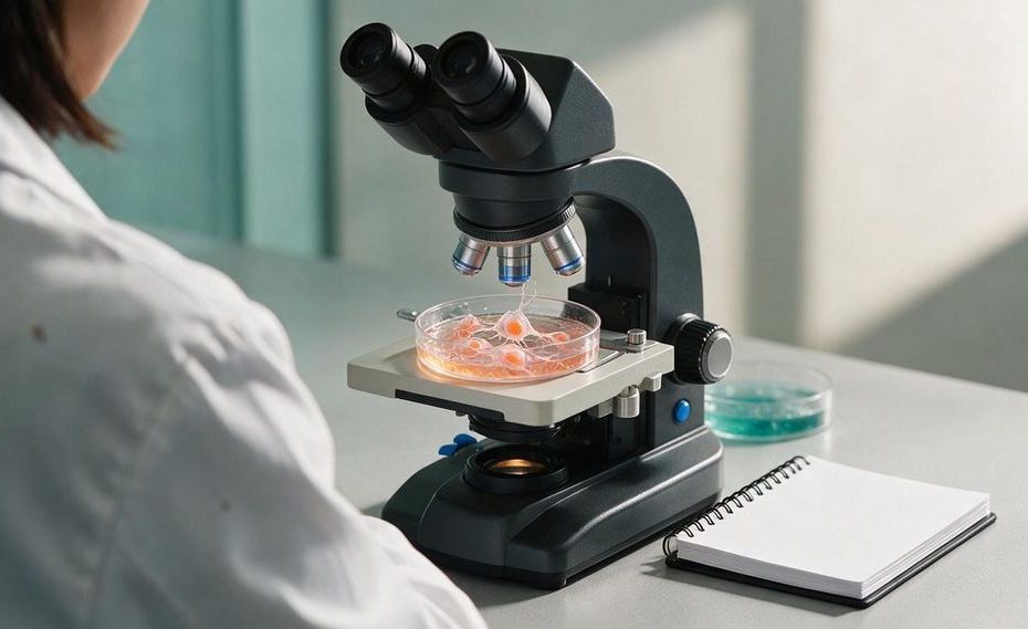
Cell adhesion is the process that lets cells attach to neighboring cells or to the material surrounding them. Cell adhesion molecules give cells the molecular grip needed to build tissues, remain anchored, communicate, move, and respond to physical and chemical signals.
Your cells aren’t isolated units. They rely on controlled attachments to form organs, guide development, repair injuries, and direct immune cells to the right location. Understanding these attachments helps you see why a failure in one adhesion protein can affect skin strength, immune defense, wound healing, or cancer spread.
What Is Cell Adhesion?
Cell adhesion describes the contacts a cell forms with another cell or with the extracellular matrix (ECM). The ECM is the network of proteins and other materials outside cells that provides structural support and biochemical signals. Adhesion connects cells to that network and to one another. The result is organized tissue rather than a loose collection of individual cells.
You can think of adhesion as a controlled molecular handshake. Proteins on a cell’s surface recognize specific partners, bind to them, and connect the attachment site to structures inside the cell. These bonds can be strengthened, weakened, assembled, or removed as conditions change. A cell can remain firmly anchored in a tissue, release part of its grip, or create new attachment points when it needs to move.
Cell adhesion isn’t the same as a cell junction. Adhesion is the broader process of attachment, and a junction is an organized structure built from adhesion proteins and supporting components. The ECM is also separate from adhesion itself because it supplies many of the surfaces and molecules that cells bind. These three ideas overlap, but they describe different parts of the same biological system.
Why Do Cells Need To Stick Together?
Cells need to stick together so they can create stable tissues, share information, and coordinate their behavior. Without controlled adhesion, cells couldn’t maintain the organized layers, boundaries, and internal structures that organs require. Your skin, intestinal lining, muscles, and blood vessels all depend on cells maintaining the right contacts. Those contacts also help tissues withstand stretching, pressure, and friction.
Adhesion gives a cell information about where it is and what it should do. When adhesion proteins bind, they can send signals into the cell that affect survival, growth, movement, and gene activity. A cell can use these signals to detect whether it has reached the correct tissue or whether the surface beneath it is stiff, soft, damaged, or changing. This conversion of physical contact into cellular signals is called mechanotransduction.
Attachment must also remain adjustable. During embryonic development, groups of cells change partners and move into new arrangements as tissues take shape. During wound healing, cells near an injury loosen selected contacts, move across the damaged area, and rebuild stable attachments. Permanent adhesion would prevent this movement, but weak or poorly controlled adhesion would leave tissues unable to hold their form.
What Is The Difference Between Cell-To-Cell And Cell-To-Matrix Adhesion?
Cell-to-cell adhesion joins neighboring cells, and cell-to-matrix adhesion connects cells to the extracellular matrix. These attachment types use different protein combinations and serve related structural and signaling functions. Cell-to-cell contacts help create continuous tissue layers. Cell-to-matrix contacts anchor those layers to their underlying support.
Cell-to-cell adhesion is common in epithelial tissues, which cover body surfaces and line internal spaces. Adhesion proteins on one cell bind to compatible proteins on the cell beside it. Inside each cell, supporting proteins connect the adhesion site to the cytoskeleton, the internal network that helps the cell maintain its shape. This arrangement lets many cells share mechanical stress rather than forcing one cell to absorb it alone.
Cell-to-matrix adhesion connects surface receptors to ECM proteins, including collagen, laminin, and fibronectin. These contacts help cells stay in place, pull against their surroundings, and migrate along a surface. They also let cells evaluate the chemical and mechanical properties of nearby tissue. Your cells use information from cell-to-cell and cell-to-matrix attachments together when deciding whether to remain anchored, divide, change shape, or move.
What Are The Main Cell Adhesion Molecules?
Cell adhesion molecules (CAMs) are surface proteins that mediate binding between cells or between a cell and the extracellular matrix. Four major families are cadherins, integrins, selectins, and members of the immunoglobulin superfamily (IgSF). They differ in what they bind, where they operate, and how long their attachments last. A tissue often uses several families at once.
Cadherins mainly support cell-to-cell adhesion and depend on calcium to maintain their binding structure. Many classic cadherins bind to the same cadherin type on a neighboring cell, creating a matched connection. E-cadherin helps preserve the integrity of epithelial tissue by keeping adjacent cells linked. Reduced E-cadherin function is associated with epithelial-mesenchymal transition and increased cancer cell invasion.
Integrins are receptors made from one alpha subunit and one beta subunit. Humans have 18 alpha subunits and 8 beta subunits that form 24 distinct integrins, each with particular binding preferences. Integrins attach cells to ECM proteins and transmit signals in two directions across the cell membrane. Signals from inside the cell can alter integrin binding, and contact with the ECM can trigger changes inside the cell.
Selectins create brief attachments that help white blood cells slow down and roll along blood vessel walls. This rolling gives immune cells an opportunity to detect local signals and prepare to leave the bloodstream. Immunoglobulin superfamily molecules participate in immune cell attachment and many other recognition events. Unlike classic cadherins, numerous IgSF adhesion proteins don’t require calcium to function.
How Do Cells Build Strong Anchors?
Cells build strong anchors by grouping adhesion proteins into organized junctions and linking those junctions to the cytoskeleton. A single molecular bond may be limited, but a cluster of bonds can distribute force across a wider area. Supporting proteins reinforce the connection and help transmit signals. The structure of an anchor depends on whether the cell is binding another cell or the ECM.
Adherens junctions use cadherins to join neighboring cells and connect those contacts to actin filaments. Desmosomes create strong cell-to-cell attachments linked to intermediate filaments, helping tissues resist mechanical strain. Hemidesmosomes anchor epithelial cells to the basement membrane beneath them. Their name reflects their appearance, but they are specialized cell-to-matrix anchors rather than half of a desmosome.
Focal adhesions are integrin-based complexes that connect the ECM to actin filaments. They often form where a cell grips a surface and generates pulling force. Proteins within these complexes help the cell sense tension and adjust its shape, movement, or internal signaling. Focal adhesions can grow when force increases and break down when the cell needs to create a new attachment elsewhere.
How Researchers Measure Cell Adhesion
Researchers study adhesion by observing how cells attach, spread, and move on controlled surfaces. Microscopy can reveal where adhesion proteins gather and how junctions change as cells move or experience force. Laboratory tests can compare how firmly cells remain attached under different conditions. Researchers can also alter a particular protein and examine how the change affects tissue organization, movement, or signaling.
How Does Cell Adhesion Support Development, Immunity, And Wound Healing?
Cell adhesion helps organize cells during embryonic development, directs immune cell movement, and supports tissue repair. In all three processes, cells must balance stable attachment with controlled release. They need enough grip to generate force and maintain order, yet they must also rearrange their contacts. This balance allows tissues to form, defend themselves, and recover from damage.
During development, cells recognize suitable neighbors and sort into organized groups. Changes in cadherin expression can alter which cells remain together, helping establish tissue boundaries and structures. Integrin attachments let developing cells interact with the ECM as they move and change shape. Adhesion signals also help cells respond to their position within a growing tissue.
During an immune response, selectins help white blood cells tether to and roll along blood vessel walls. Stronger adhesion then allows selected cells to stop and pass into nearby tissue. This sequence places immune cells where they are needed instead of leaving them to circulate without direction. Defects in the required adhesion proteins can interfere with immune cells reaching an infection.
Wound healing also depends on timed changes in adhesion. Cells at the edge of an injury adjust existing junctions, attach to the exposed matrix, and move across the damaged area. New cell-to-cell contacts help restore the tissue barrier once the gap closes. Poor adhesion can slow this process, and adhesion that remains too rigid can interfere with cell migration.
What Happens When Cell Adhesion Fails?
Failed or altered cell adhesion can weaken tissue, disrupt immune defense, delay healing, and help abnormal cells move into new locations. The outcome depends on which protein is affected and where that protein normally works. A defect in an epithelial anchor can cause tissue separation. A defect in an immune cell integrin can prevent white blood cells from leaving the bloodstream efficiently.
Leukocyte adhesion deficiency (LAD) is a rare inherited immune disorder associated with defects in adhesion proteins, including beta-2 integrins. White blood cells have trouble attaching properly and moving from the blood into infected tissue. Common features include recurrent bacterial infections, poor wound healing, and delayed separation of the umbilical cord. These problems show that immune defense requires movement and controlled attachment rather than circulation alone.
Epidermolysis bullosa is a group of inherited conditions that cause fragile skin and blistering after minor friction. Some forms involve defects in proteins that connect epithelial cells to the basement membrane or support hemidesmosome function. When these anchors can’t withstand mechanical stress, tissue layers separate more readily. The resulting damage demonstrates how cell-to-matrix adhesion protects tissues during ordinary movement and contact.
Cancer can also involve altered cell adhesion. Changes in cadherins and integrins can help tumor cells loosen their contacts, interact differently with the ECM, migrate, and establish growth in other tissues. Loss of E-cadherin is associated with changes that support epithelial cell invasion. Adhesion changes don’t act alone, but they contribute to several steps involved in metastasis.
Stronger adhesion isn’t always better. Cells must form the right attachment, in the right place, for the right amount of time. An attachment that can’t release may block migration, and one that forms in the wrong tissue may disrupt normal organization. Healthy cell adhesion depends on precise regulation rather than maximum stickiness.
Why Is Cell Adhesion Important?
- Holds cells together to form tissues and organs
- Anchors cells to the extracellular matrix
- Guides development and wound healing
- Lets cells sense mechanical and chemical signals
- Helps immune cells migrate and stick where needed
Cell Adhesion Turns Contact Into Coordinated Action
Cell adhesion gives your cells far more than a place to hold on. It lets them build stable tissues, read physical signals, communicate with neighboring cells, move through the body, and repair damaged areas. Cadherins, integrins, selectins, and immunoglobulin superfamily proteins manage different parts of this work, often through organized junctions connected to the cytoskeleton. When cell adhesion molecules are missing or poorly regulated, the effects can appear in fragile skin, impaired immunity, delayed healing, or cancer invasion. Your tissues remain organized because cellular attachments are strong when support is needed and adjustable when cells need to move.
References
- National Center for Biotechnology Information Bookshelf: Cell Junctions, Cell Adhesion, and the Extracellular Matrix
- Encyclopaedia Britannica: Cell Adhesion
- National Cancer Institute Dictionary: Cell Adhesion Molecule
- PubMed: Integrins, Bidirectional Allosteric Signaling Machines
- MedlinePlus Genetics: Leukocyte Adhesion Deficiency
- National Center for Biotechnology Information GeneReviews: Leukocyte Adhesion Deficiency
- Genetic and Rare Diseases Information Center: Epidermolysis Bullosa.

Nirdosh Jagota is Managing Partner at GRQ Biotech Advisors with 30+ years in the biotech industry. A former executive at Amgen, Genentech/Roche, Merck, and Pfizer, he has led >25 NDAs/BLAs/MAAs and hundreds of INDs across global regulatory, quality, and compliance.