
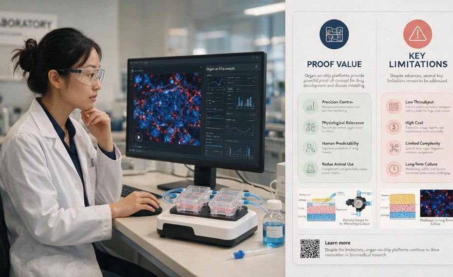
Organ-on-chip models can prove specific human tissue responses under controlled conditions, including barrier behavior, mechanical-force effects, and certain toxicity signals. Their organ-on-chip limitations become apparent when a study depends on whole-body immunity, circulation, endocrine feedback, long time scales, or reproducible scale-up.
These systems can narrow the translational gap between conventional cell culture, animal testing, and human trials. Yet a chip remains a simplified functional unit, not a miniature human organ. Understanding that distinction helps you choose where microphysiological systems can strengthen a development program and where additional models remain necessary.
Why Does Preclinical Translation Fail So Often?
Preclinical translation often fails because a model can produce internally consistent results without reproducing the human biological process that matters. Static two-dimensional cell culture removes many physical forces, tissue interfaces, and fluid conditions found in a living organ. Animal models restore whole-body biology, but species differences can still hide human toxicity or overstate a treatment effect.
The translational gap becomes costly when a candidate passes early safety studies and fails after development has advanced. Late failure means that earlier experiments answered the wrong biological question, used a model with limited human relevance, or measured an endpoint that didn’t predict clinical performance. Organ-on-chip models are designed to reduce that risk by placing human cells in controlled microenvironments that reproduce selected tissue functions.
You should treat the chip as a decision filter, not a universal proof engine. It can help you reject a weak candidate sooner, compare lead compounds, study a suspected toxicity mechanism, or test how physical forces change a tissue response. It cannot establish whole-body safety simply because its cells are human.
What Can Organ-on-Chip Models Prove Better Than Conventional Models?
Organ-on-chip models perform best when the research question involves a defined tissue interface, mechanical force, flow condition, or organ-specific response. Microfluidics can expose cells to controlled flow, concentration gradients, stretch, and contact between different cell types. These conditions offer more physiological relevance than a flat culture plate for selected mechanisms.
The original lung-on-chip research showed why mechanical design matters. Researchers recreated an air-facing tissue layer, a blood-facing layer, and cyclic stretch associated with breathing. Applying that stretch changed the inflammatory response to silica nanoparticles, demonstrating that a physical force could alter a biological outcome that static culture might miss.
Liver toxicity provides another strong use case. A human Liver-Chip assessment correctly identified 87% of the tested drugs known to cause drug-induced liver injury (DILI) in people after those drugs had passed animal testing. That result doesn’t prove that every liver chip will predict every human reaction, but it shows that a validated human tissue model can detect specific hazards missed by established preclinical methods.
Where Do Organ-on-Chip Limitations Become Biologically Important?
The main organ-on-chip limitations emerge when the biological outcome depends on systems that extend beyond one tissue compartment. A chip may reproduce epithelial barrier function, local inflammation, or organ-specific toxicity without recreating the full adaptive immune system. It may include selected immune cells, but that is different from modeling immune development, circulation, recruitment, memory, and coordinated responses across several organs.
Functional vascularization creates a related problem. Flow through a manufactured channel can mimic parts of blood movement, yet it doesn’t automatically reproduce a living vascular network that grows, remodels, leaks, repairs itself, and exchanges signals with surrounding tissues. Long-term tissue survival also depends on reliable delivery of oxygen and nutrients, waste removal, and stable cellular organization.
Chronic and systemic conditions raise the difficulty further. Endocrine loops can involve several organs releasing signals at different concentrations and time intervals. Neural regulation, metabolism, immune activity, and changing tissue states may influence the same outcome. A single-organ chip can isolate one part of that process, but it cannot prove how the entire process behaves inside a person.
Why Can Chip Materials Distort Drug Data?
The material surrounding the cells can influence the result as much as the biological design. Polydimethylsiloxane (PDMS) remains common in microfluidic devices because it supports rapid fabrication and flexible channel designs. Its tendency to absorb small hydrophobic molecules, including many drugs, creates a serious measurement problem.
If PDMS removes part of a compound from the circulating medium, the cells receive a lower exposure than the nominal dose suggests. Your recorded concentration may say one thing, yet the biologically available concentration may be different. Dose-response curves, toxicity thresholds, and pharmacokinetic estimates can then shift without an obvious experimental warning.
Alternative materials include polystyrene and cyclic olefin copolymer, but changing the material doesn’t remove the need for validation. You still need to measure compound recovery, adsorption, evaporation, channel compatibility, and cellular response under the actual assay conditions. Material selection should be part of the experimental plan, not a manufacturing detail considered after the device has been chosen.
Why Is a Body-on-a-Chip Still So Difficult to Build?
A body-on-chip connects several tissue compartments so that metabolites and signals can move between them. Current advanced systems can link three or four organ compartments, commonly representing tissues from the liver, heart, lung, or kidney, for periods measured in weeks. Moving from a few connected tissues to ten physiologically balanced organs remains an unresolved engineering problem.
Every organ has different nutritional, mechanical, and chemical requirements. A medium that maintains liver cells may not support heart, kidney, and lung cells equally well. Connecting the compartments can also dilute signals, create unwanted flow conditions, or expose one tissue to concentrations that don’t match human physiology.
Organ ratios create another barrier. A full-size liver, kidney, heart, and lung don’t contribute equal tissue mass, blood volume, metabolic capacity, or clearance. Shrinking them into chip compartments requires more than making every chamber smaller by the same amount. You must balance tissue size, flow, exposure, and metabolic output so that one compartment doesn’t dominate the entire system.
Does the Food and Drug Administration Accept Organ-on-Chip Data?
The Food and Drug Administration (FDA) can consider organ-on-chip data, but acceptance doesn’t mean automatic approval or a universal replacement for animal studies. Federal law no longer requires every new drug to undergo animal testing before human trials. Alternative methods, including organ-on-chip platforms and other microphysiological systems (MPS), can now contribute to the evidence supporting development decisions.
The intended use still matters. A platform validated for detecting a defined liver toxicity signal doesn’t automatically qualify for cardiac safety, kidney clearance, immune response, or systemic exposure. Regulators need evidence that the model is reliable, reproducible, and suitable for the decision attached to its data.
You should define that decision before designing the study. A chip may support mechanism testing, candidate ranking, hazard identification, dose selection, or supplementary safety evidence. Claims that extend beyond the model’s validated purpose weaken the submission rather than strengthening it.
How Should You Use Organ-on-Chip Models in Drug Development?
Use organ-on-chip systems where human-specific tissue behavior can change a development decision. They fit high-content evaluation of selected lead candidates better than primary screening of very large compound libraries. The added biological detail is most valuable after simpler assays have narrowed the candidate pool.
Start with a precise question and choose the chip around that question. Define the relevant cell types, flow conditions, exposure route, mechanical forces, extracellular matrix, assay duration, and measurable endpoints. Record cell seeding density, medium composition, flow rate, device material, and acceptance criteria so another laboratory can reproduce the work.
You can also pair chip data with conventional cell culture, animal evidence, and computational modeling. Each method answers a different part of the safety or efficacy question. Agreement across independent methods strengthens a conclusion, and disagreement can reveal where additional testing is needed.
What Must Improve Before Organ-on-Chip Models Can Scale?
Standardization must reduce variation between laboratories, device batches, cell sources, and operating procedures. Differences in flow rate, cell maturity, medium formulation, extracellular matrix, and endpoint selection can produce conflicting results from devices carrying the same organ label. Shared reporting practices would make it easier to compare studies and identify whether a failed result came from the compound or the platform.
Automation can improve fluid handling, exposure timing, sampling, and data capture. It can also reduce operator variation and support larger experiments without stripping away the biological features that make the chip useful. Scale should be measured through decision quality and reproducibility, not only through the number of wells processed.
Induced pluripotent stem cells (iPSCs) offer a route to patient-linked research, but their maturity and consistency require careful review. They may help you study genetic differences or compare responses among cell donors when the model has a well-defined endpoint. Their human origin doesn’t remove the need to confirm tissue function, phenotype stability, and assay performance.
What Are the Main Organ-on-Chip Limitations?
- Missing a complete adaptive immune system
- Limited functional vascularization
- Drug absorption by chip materials
- No universal reproducibility standards
- Weak modeling of chronic multi-organ loops
Use Organ-on-Chip Evidence for the Question It Can Answer
Organ-on-chip models can reveal human tissue responses that flat culture and some animal studies miss, particularly when flow, barriers, mechanical force, or organ-specific toxicity shapes the outcome. Their value comes from proving a defined mechanism under controlled conditions, not from recreating an entire person. You’ll get stronger evidence when you match the device to a narrow decision, measure material effects, document operating conditions, and compare the result with other models. The most practical response to organ-on-chip limitations is disciplined use: rely on the technology as a human-relevant filter, then add broader systems when immunity, circulation, endocrine signaling, or whole-body exposure controls the answer.
References
- Communications Medicine: Performance Assessment and Economic Analysis of a Human Liver-Chip for Predictive Toxicology
- Wyss Institute: Human Organs-on-Chips
- National Center for Advancing Translational Sciences: Tissue Chip for Drug Screening
- Food and Drug Administration: Advancing Alternative Methods
- Lab on a Chip: Small-Molecule Absorption by Polydimethylsiloxane
- PubMed: Small Molecule Absorption by Polydimethylsiloxane in Drug Response Assays
- Science: Reconstituting Organ-Level Lung Functions on a Chip
- International Consortium for Innovation and Quality in Pharmaceutical Development.

Nirdosh Jagota is Managing Partner at GRQ Biotech Advisors with 30+ years in the biotech industry. A former executive at Amgen, Genentech/Roche, Merck, and Pfizer, he has led >25 NDAs/BLAs/MAAs and hundreds of INDs across global regulatory, quality, and compliance.